MAIT Immune Cell Findings Unite U.S. and U.K. ME/CFS Researchers
The US and the UK are said to have a "special relationship". That special relationship hasn’t generally extended to ME/CFS research, given a decidedly different focus on ME/CFS in the two countries - a strong focus on biological research in the US and more of a focus on CBT/GET in the UK. That might be changing, though.Derya Unutmaz at Jackson Labs and Jacqueline Cliff of the London School of Hygiene and Tropical Medicine | LSHTM appear to have both independently landed on the same immune cell in chronic fatigue syndrome (ME/CFS). Given the multitude of immune cells found in the body, that has the potential to be rather special. The specialness doesn't stop there. The samples tested by these two teams - all 300 of them - come from the UK ME/CFS Biobank - which since 2014 has received major funding from the National Institutes of Health (NIH) in the US. (The Biobank has also received funding from The ME Association, Action for ME, and ME Research UK.) Plus, the NIH provided most of the funding for the Cliff project.The UK ME/CFS Biobank is big. It contains serum, plasma, peripheral blood mononuclear cells (PBMC), red blood cells/granulocyte pellet, whole blood, and RNA samples from over 500 ME/CFS and multiple sclerosis patients and healthy controls. Plus, it includes an extensive dataset of 700 clinical and socio-demographic variables.The Cliff study focused on the immune system - a natural system to target given the infectious onset many experience and the symptoms common to all patients. An immune "hole" could give a pathogen time to do more damage, set off an autoimmune response, or alter immune functioning in some other way.Immune studies in ME/CFS are not uncommon, but the Cliff team researchers (sounding very English at least to my ears) described their results as "discrepant" and inconclusive. Interesting research findings have not been reproduced in ME in part, they asserted, because of small study sizes, varied research methods, and sometimes a less than stellar quality of the studies.This Biobank study is different, they believe. A large study with a well characterized patient group, they clearly believe its results will stand the test of time.
The Study
Front Immunol. 2019 Apr 16;10:796. doi: 10.3389/fimmu.2019.00796. eCollection 2019. Cellular Immune Function in Myalgic Encephalomyelitis/Chronic Fatigue Syndrome (ME/CFS). Cliff JM1, King EC1, Lee JS1, Sepúlveda N1,2, Wolf AS1, Kingdon C3, Bowman E3, Dockrell HM1, Nacul L3, Lacerda E3, Riley EM1.
The Patients
The Cliff study examined the samples from over 400 patients and controls (251 ME/CFS (54 severely affected and 197 mild/moderate), 46 multiple sclerosis, 107 healthy controls.)The patients met either the Canadian Consensus Criteria or the 1992 Fukuda Criteria (or both) and were mostly recruited via the UK National Health Service. The patients were determined to meet either criteria after their responses to a Symptoms Assessment form were fed into a computerized algorithm that maps their symptoms onto the different ME/CFS study case definitions.Since the Fukuda definition does not require post-exertional malaise - the core symptom of ME/CFS - it was surprising to see the group potentially accept patients who only met that definition. It wasn't clear from the study what proportion of patients, if any, met only the Fukuda criteria, though. Severely ill patients were mostly home or bed bound. Their blood samples were taken during home visits.People who had taken antiviral medications or drugs known to alter immune functioning, had a recent history of vaccination, had a history of other chronic diseases such as tuberculosis, cancer, uncontrolled diabetes, etc., had a severe mood disorder, or who had been pregnant or breastfeeding in the past 12 months were excluded.One part of the study focused on natural killer (NK) cells - key players in the early, innate immune response. Given the NK findings in ME/CFS, the group's decision to analyze NK cells was not a surprise, but they gave their analysis a twist. Because cytomegalovirus (CMV) infections have such profound effects on our NK cells (and the rest of our immune system), the relationship between CMV infections and NK cells was assessed to determine whether a past CMV infection could be responsible for the NK cell abnormalities seen in ME/CFS.
Results
The Next Big Thing in Immune Research? MAIT cells Pop Out Again
The big news from the Cliff study is the increased frequency of the CD8+ mucosal associated invariant T cells or MAIT cells. The UK authors noted that an increased frequency of MAIT T-cells has not been published before, which is true, but Derya Unutmaz, leader of the NIH ME/CFS Research Center at the Jackson Labs, has been talking about them in ME/CFS for several years.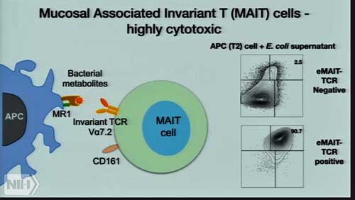 Unutmaz reported finding high levels of MAIT cells in ME/CFS patients. Unutmaz’s findings suggest that the MAIT T-cells have been repeatedly activated in ME/CFS and that they evidence the same activated/burned out pattern he’s found in other T-cells. He and Dr. Oh at Jackson Labs are trying to determine which stomach bacteria has turned them on and then find a way to eliminate or reduce it.MAIT cells are known for the role they play protecting the lining of the gut against toxic bacteria. Their name – mucosal invariant T-cells – derives from the high levels of these cells gathered around the mucosal surfaces of the gut (e.g. the lining). In effect they are the gut’s innate immune cells - sentinels guarding the gut wall which can, in contrast to other T-cells, react immediately to invaders.They’re different from other T-cells which get activated after being triggered by an antigen from a pathogen. Instead, they're activated by fats and vitamin B2 metabolites produced by plants, bacteria (E. coli, Pseudomonas aeruginosa, Klebsiella pneumonia, L. acidophilus, S. aureus, and S. epidermidis, C. albicans, C. glabrata, and S. cerevisiae ) and fungi. Because cytokines produced by viral infections can activate them as well, the high degree of MAIT cell activation is not necessarily due to bacteria in the gut - but it’s the most likely scenario.MAIT research only started popping up after 2010, when studies revealed these unusual cells were able to detect bacteria and fungi and respond with pro-inflammatory cytokines. Since then many studies have suggested that MAIT cells play an important role in infectious diseases, autoimmune diseases and cancer. MAIT cells are not always pro-inflammatory, but increased levels, particularly of cytotoxic MAIT cells, are believed to be associated with pathogenic states.In contrast to Unutmaz's apparent (but unpublished) findings of high levels of MAIT cells in ME/CFS overall, this study found a high proportion of MAIT cells only in the severely ill ME/CFS patients. They noted that a small number of the severely ill patients were reported to have "exceedingly high” frequencies of these cells.Most of the MAIT cells in the severely affected ME/CFS patients (as well as in the MS patients) were in their cytotoxic (killing) form. They'd probably been activated by a bacterium in the gut and were apparently on the prowl, ready to pounce. While the increased proportion of MAIT cells only weakly discriminated the severe ME/CFS patients from the healthy controls, the high percentage of killer T-cells (cytotoxic T-cells) found was moderately discriminative.Interestingly, the Cliff study authors pointed out that peripheral MAIT cell levels in healthy volunteers can increase 2-fold following exercise. Finding similarly high levels of MAIT cells in the severely ill patients suggested they were in a similar post-exercise state without having engaged in any exercise.
Slight Increase in ESR Surprises
Interestingly, symptoms associated with inflammation/infection were more common and more severe in the ME/CFS cohort than in the MS cohort (go figure!). Perhaps that's not a surprise, since ME/CFS has been shown to impact functioning to a greater degree than MS.The slight raise in erythrocyte sedimentation rate (ESR) - an inflammatory marker - in mild/moderate cases of ME/CFS compared to the other groups (Including the severe ME/CFS group) was surprising, though, given that very low ESR's are thought to be typical in this disease.
Laboratory studies. These tests can be used to exclude other diseases associated with fatigue. The most consistent laboratory abnormality in patients with CFS is an extremely low erythrocyte sedimentation rate (ESR), which approaches zero. Typically, patients with CFS have an ESR of 0 to 3 mm/h. A normal ESR or one that is in the upper reference range suggests another diagnosis. https://www.consultant360.com/content/chronic-fatigue-syndrome-update-diagnosis-primary-care
Natural Killer Cells
As the Cliff study introduced a new factor into ME/CFS research (MAIT cells), it took a hatchet to the last big immune finding in ME/CFS - natural killer cells. The study found no significant differences in NK cell proportions, types, KIR receptors or activation markers before or after they stimulated them.Some NK markers did stand out, but only in patients who had been exposed to CMV. The authors suggested a past CMV infection in some of the ME/CFS patients had likely caused the NK cell abnormalities in ME/CFS - not ME/CFS.The Cliff study, however, used a different test of NK cell functioning than some groups have used in the past. The British group assessed both T and NK cell functioning by determining how the cells responded to stimulation; i.e., did they produce distinctive markers and/or start producing cytokines. The ME/CFS patients’ cells apparently whizzed through that test - they perked up and started producing cytokines, leaving the authors to report that no functional issues with these cells are present. Dr. Klimas, however, uses a more direct functional NK cell assay which measures the number of target cells killed. Plus, instead of the PBMC's used in the Cliff study, she uses whole blood - possibly a critical factor, given Ron Davis' and Fluge's findings that something in the plasma is affecting the cells. In fact the first hint of a blood-borne factor in ME/CFS showed up in NK cell studies. That idea that something in the blood was impacting functioning first showed up when Dr. Klimas realized that a study which found no evidence of problems with NK cell functioning had not used whole blood in its tests.The UK study authors noted that small study sizes have hampered immune results in this disease, but size was not an issue for the 2011 Klimas/Fletcher study (176 ME/CFS patients, 230 healthy controls) which found significant declines in NK cell functioning, and those declines were associated with increased fatigue levels. In an Australian study, Brenu also used a target cell killing test to show reductions in T-cell functioning. The UK study authors did not allude to other possible functional tests or the whole blood issue in their manuscript.
Exhausted T-cells?
The UK study authors did find a number of T-cell abnormalities: increased proportions of effector memory CD8+ T cells, decreased proportions of terminally differentiated effector TEMRA cells, and some minor changes elsewhere - whose effects are unclear. The UK authors suggested, though, they could be due to "ongoing antigenic stimulation" due to an unresolved infection or autoimmunity.Either could presumably produce a state of "immune exhaustion" which some have hypothesized is present in ME/CFS.Derya Unutmaz focused on key players in autoimmunity and inflammation called TH17 cells in his U.S. study. He wasn’t surprised to find high levels of TH17 cells - which are regulated by the gut - but he was shocked to find low levels of the IL-17 cytokine they produce. That finding also suggested that the immune cells in ME/CFS might be in a state of exhaustion.The Cliff study's IgG antibody tests found no evidence of increased herpes virus reactivation in ME/CFS, and some evidence of it in MS. The group didn't close the book on the possibility of herpesvirus reactivation in ME/CFS, though, stating that other antibody tests for EBV might produce different results.
Conclusion
The Cliff study was a large UK Biobank study using both Fukuda and/or Canadian Consensus Criteria to identify its patients. The study's finding of moderately increased ESR levels in the mild/moderate patients was surprising, given past reports of low ESR levels in ME/CFS.Except in patients who have been exposed to cytomegalovirus (CMV) in the past, the study found no evidence of natural killer cell issues in ME/CFS. The researchers did not, however, use a functional assay used successfully in the past which more directly measures NK or T-cell killing capacity.The study's major finding was a significant increase of specialized T-cells called MAIT cells in the severely ill patients. MAIT cells are found across the body but are most known for the role they play protecting the gut lining from toxic bacteria. High levels of MAIT cells have been associated with infectious diseases, autoimmunity and cancer.This is the second recent and, it should be noted, independent report of high levels of MAIT cells in ME/CFS. In fact, these two reports are the first time MAIT cells have been implicated in this disease.Dervy Unutmaz and Dr. Oh of the Jackson Labs are currently trying to isolate the bacteria triggering the high levels of MAIT cells they've found in ME/CFS.The Cliff study did find moderate T-cell anomalies which could possibly reflect a state of chronic T-cell activation caused by an infection or autoimmune response. Derya Unutmaz also recently reported he'd found evidence of immune cell exhaustion in his T-cell studies.