ME/CFS Seahorse Energy Production Study Shows Surprises
Dr. Maureen Hanson leads one of the three NIH funded ME/CFS research centers, but her ME/CFS research doesn't stop there. Using samples from Dr. Daniel Peterson provided by the Simmaron Research Foundation, she's also been assessing the metabolism of one of the most important cells in our immune systems: our T-cells.T-cells affect a large part of our adaptive immune response that clears out infections. They do this by regulating our immune response (CD-4 or Helper T-cells) and/or by killing off pathogens that have infected other cells (CD-8 or cytotoxic T-cells).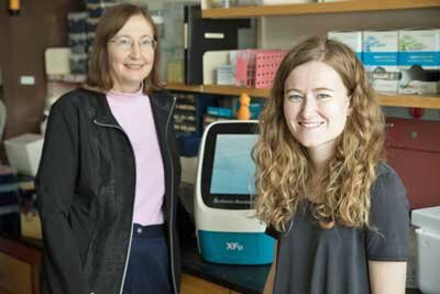 Prior to getting activated, T-cells are primarily on sentry duty. Once activated by dendritic cells presenting little bits of pathogens to them things change dramatically, however. The T-cells rev up their cellular engines to order to start pumping out cytokines or clones en masse in order to stop the infection. Both parts of energy production - glycolysis and oxidative phosphorylation - have to jump into action.In short, assessing the energy production of activated T-cells is a perfect way to determine if their energy metabolism has been affected in ME/CFS - and that’s just what Maureen Hanson's group did.Alexandra Mandarano, a graduate student in Hanson's lab, took T-cells from 53 healthy controls and 45 pretty long duration (avg. a@ 12.7 years) ME/CFS patients and healthy controls and tricked them into going into high alert with antibodies plus IL-2. Then, using the Seahorse Flux Analyzer, she examined how well the two parts of their cellular energy production system did in both unactivated and activated T-cells: glycolysis - the anaerobic part which takes place outside the mitochondria, and oxidative phosphorylation - the aerobic part which takes place inside the mitochondria (and produces far more ATP) .
Dr. Hanson presented on her results at the recent Open Medicine Foundation sponsored Harvard Symposium
https://www.youtube.com/watch?v=QAdZNU6D7Gs
Results
Whether they were activated or not, mitochondrial energy production; i.e. oxidative phosphorylation (the main ATP producer) was normal for both the CD4 and CD8 cells in the ME/CFS group. When pushed, the mitochondria in the ME/CFS patients' cells quickly got energy production up to speed. That was a surprise. Usually when you push a cell or system in ME/CFS it fails- but, in this case, the T-cells responded normally.Then came the real surprise. Systems in ME/CFS often test out fine or at least not strongly abnormal at baseline or rest, but in this case Hanson found low glycolysis activity in both the T-helper cells (CD4) and the CD8 cells at baseline. Simply prowling around the body, they had considerably lower levels of glycolytic activity. When pushed, though, their glycolytic activity was normal. The pattern was opposite to what we usually see.That wasn't all. It's possible with the Seahorse to turn off different energy production pathways in order to assess how effectively the other pathways are at compensating. When the oxidative phosphorylation pathways were turned off experimentally, the ME/CFS patients' glycolytic pathways failed to compensate as effectively as did those of healthy controls. Thus no problems with mitochondrial energy production were found but three potential issues with glycolysis popped up: low glycolytic activity in both forms of unactivated T-cells, and poor glycolytic compensation with the oxidative phosphorylation pathways were turned off.Hanson’s group next examined a critical part of energy production called the mitochondrial membrane potential. Our mitochondria need to maintain a certain membrane potential to keep up the flow of positively charged ions into the mitochondria. It does this by keeping more positively charged ions outside of the mitochondria and more negatively charged ions inside the mitochondria. Her group used a flow cytometer to assess the levels of mitochondria present and to determine how strong the membrane potential was.The mass and membrane potential of the ME/CFS patients' CD4 T-cells and the mitochondrial mass of the CD8 cells was normal, but the membrane potential of the CD8 T-cells - whether activated or not - were significantly impaired in the ME/CFS patients.Four potential problems, then, were found:
- low glycolytic activity in unactivated CD4 and CD8 T-cells
- poor glycolytic compensation when the oxidative phosphorylation pathways were turned off
- The mitochondrial membrane potential was impaired in the CD8 T-cells
Dr. Hanson left her presentation with the encouraging statement that we are starting to put the pieces of the puzzle together in ME/CFS and the tantalizing suggestion that ME/CFS might be something different than what we think it is right now; i.e. keep an open mind, don't put all your eggs in one basket, and be prepared for surprises.
Overview
Hanson and her co-authors have submitted a paper and we will get more details when their paper is published but, with these preliminary results, we have a few more data points on cellular energy production in ME/CFS. While noting that several study results are pending, maybe it's time for a look at what we have.It should be noted that measuring energy production is very complex. Different researchers are doing it in different ways, and I am no judge of any of them. Researchers are using different instruments, different criteria, different kinds and numbers of patients, and they are reporting things differently. Solving those problems is one of the reasons for the NIH funded ME/CFS research centers where larger studies can use proven technologies and rigorously defined patient populations.Check out some of the different protocols which have assessed mitochondrial functioning in isolation from the blood in ME/CFS:
Study protocols
- Hanson's group activated her T-cells using antibodies and IL-2 and then tested activated and unactivated cells in the Seahorse Machine
- Tomas took PBMC's and stressed them in the Seahorse machine
- Stanford took PBMC's and then used laboratory assays to test each of the complexes and flow cytometry to assess mitochondrial membrane potential
- Fisher (unpublished) appears to have taken PBMC's and stressed them in the Seahorse machine
- Vermeulen measured ATP PBMC's etc. in the lab
- Smits measured ATP production rate in muscle biopsies
The Land of Mixed Signals
We seem to find ourselves in a familiar place - the land of mixed signals! One encouraging unmixed signal is that everyone seems to be finding something wrong - just often different things.
MITOCHONDRIA
Mitochondria Mass - Normal
- Hanson - CD4 and CD8 (T-helper cells)
- Fisher
Mitochondria ATP production - Normal
- Hanson (T-cells)
- Stanford study (not a Ron Davis study) (PBMC's)
- Fisher (PBMC's)
- Vermoulen (PBMC's)
- Smits (muscle biopsy)
Increased ATP Production Overall
- Stanford
- Preliminary results from NIH Intramural study
Reduced ATP production
- Tomas - under both low and high glucose conditions
Functioning of Complexes - Normal
- Stanford (I-IV)
- Vermeulen (I-II)
Functioning of Complex V - reduced
- Fisher
GLYCOLYSIS
Increased Glycolysis at Baseline (PBMC's)
- Stanford
Reduced Glycolysis at Baseline (T-cells)
- Hanson
Reduced Glycolysis (low glucose conditions)
- Tomas
Reduced Compensatory Glycolysis
- Hanson
- Tomas (?)
Glycolysis Stress Test (Glycolysis, glycolytic reserve, glycolytic capacity)- normal
- Tomas
- Fisher
It's quite a muddle. Surprisingly, though, the most consistent finding thus far is normal (or in two cases) increased mitochondrial production (!) Not many studies have directly measured glycolysis, but in these early days the results are mixed.
Isolation
 Note that all these studies are assessing the energy production of the mitochondria in isolation. None tested cells in the blood where Davis, Fluge and Mella and Prushty have found evidence that some sort of inhibiting factor may be present. The metabolomic findings which suggest problems with glycolysis are present have been assessing factors in the blood and urine as well.Adding an exercise stress test would, of course, add another important factor. At the NIH ME/CFS Conference, Brian Walitt reported that the NIH is finding that exercise causes mitochondrial oxygen consumption (ATP production) to increase in the healthy controls but to decrease in about half of the ME/CFS patients. Several recent studies have validated that exercise impairs energy production in ME/CFS (blog coming up). Where and how the energy depletions are occurring is unclear. (Note that most of these studies examined immune cells not muscle cells.)We obviously have long way to go to fit all the different pieces of the energy production puzzle in ME/CFS together but the good news is that an increasing amount of research is now being aimed at deciphering what's inhibiting energy production in this disease.The Simmaron Research Foundation's collaboration with Maureen Hanson - which paired rigorously diagnosed patients with a respected researcher - is just one way the Foundation is contributing to solving that puzzle.